Advanced microscopy
Raman microscopy (CARS & SRS)
Coherent anti-Stokes Raman scattering (CARS) and stimulated Raman scattering (SRS) are used for high-speed and high-sensitivity label-free vibrational imaging.
Introduction
What CARS and SRS add
Vibrational imaging provides chemical specificity, which is useful in biological microscopy, and pharmaceutical and material science, especially where other microscopy techniques provide ambiguous results and labels cannot be used. Spontaneous Raman scattering microscopy, while sensitive, is slow. However, its nonlinear counterparts – coherent anti-Stokes Raman scattering (CARS) and stimulated Raman scattering (SRS) – can be used for high-speed and high-sensitivity label-free vibrational imaging.
How CARS and SRS differ
In CARS microscopy, two beams are used to produce radiation at a third wavelength via a vibrational level of the sample. The energy difference between the two beams can be tuned to different vibrational levels, thus providing chemical imaging contrast. The produced CARS signal is at a different wavelength that is usually background-free, which makes its detection relatively simple. However, CARS distorts the vibrational lineshapes and possesses a nonresonant signal component, which complicates chemical attribution. SRS is free from these disadvantages; however, its signal is a small modulation on one of the incoming beams and requires an elaborate detection scheme.
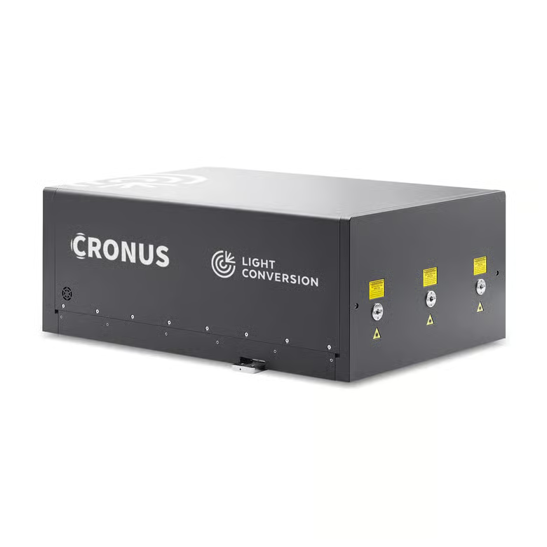
CRONUS-2P for dual-beam Raman
CRONUS-2P is a femtosecond laser providing three simultaneous and synchronized outputs with high repetition rate, high output power, short pulse duration, and GDD control. Two outputs are independently tunable in the 680–960 nm and 940–1300 nm ranges, while the third is fixed at 1025 nm. The three simultaneous outputs enable advanced CARS and SRS applications with dual-band imaging, a broader selection of vibrational resonance frequencies, constant-difference dual-beam tuning, and resonant enhancement.
SRS of polymeric materials
(a) Spontaneous Raman and SRS spectra of polymeric materials, normalized with the molecule of interest maximum peak. (b) SRS images of dyed and un-dyed PS beads. Images were recorded using CRONUS-2P.
SRS of cells and β-carotene
(a) Imaging of nuclei of AZ568 stained, fixed HeLa cells with SRS (red) and fluorescence (green). (b) SRS and spontaneous Raman spectrum (at 488 nm) of 1 mM solution of AZ568 in DMSO-d6. (c) Structure of β-carotene. (d) β-Carotene spectrum recorded with SRS and spontaneous Raman. (e) Spectral focusing spectrum of β-carotene solution in DMSO-d6. The images were recorded using CRONUS-2P.