
AI-Assisted Analysis for Laser Microscopy Time Series: Evidence, Workflows, and Frontier Directions
An evidence-based guide to reviewing laser-microscopy time series, including three-photon, CARS/SRS, transient-absorption, and ultrafast-electron workflows.
Laser-microscopy time series can be scientifically powerful, but the result is only as defensible as the connection between acquisition settings, image-processing choices, measurements, and review. AI can assist with transparent analysis code; it does not turn a catalogue specification, a published result from another system, or an unvalidated segmentation into a conclusion.
Start by separating three kinds of evidence
An instrument catalogue describes a configuration. A paper describes a particular optical system, sample, and validation protocol. An analysis tool describes a downstream workflow. Those are three different evidence layers, and none should be silently used as proof of another.
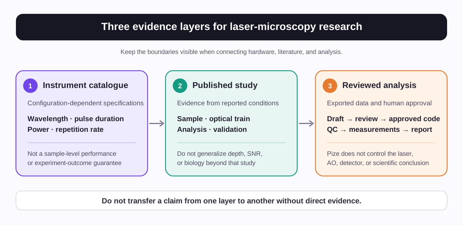
- Catalogue specification: wavelength range, pulse duration, repetition rate, and power are manufacturer-published values for a named configuration. They are not sample-level performance guarantees.
- Published workflow: a paper can establish what happened in its reported system and conditions. Its depth, signal-to-noise ratio, or biological result should not be generalized beyond that evidence.
- Reviewed downstream analysis: code may organize, quantify, and report exported data, but the research team must validate preprocessing, models, controls, and interpretation.
This distinction is especially important for time-lapse work, where a visually convincing movie can conceal registration drift, photobleaching, segmentation bias, incomplete metadata, or an invalid statistical unit.
A reviewable workflow for microscopy time series
The four familiar stages—registration, segmentation, tracking, and quantification—remain useful. A rigorous workflow also makes the acquisition and validation steps explicit.
| Step | What the research team should record or test | A defensible role for Pize after data export |
|---|---|---|
| Acquisition ledger | Sample, channel, voxel size, objective, timestamps, excitation wavelength, power at the sample where measured, and relevant system settings | Help draft a structured metadata table and a data-quality checklist |
| Registration QC | Fiducials or stable structures, residual motion, interpolation choices, and a before/after visual check | Help draft a small, reviewable registration-QC script and plots |
| Segmentation validation | A held-out set with annotations or expert review, failure cases, and thresholds | Help compare a proposed method and generate a validation report; it does not certify a model |
| Tracking and measurement | Link parameters, gaps, split/merge policy, units, and the biological unit of replication | Help make trajectory tables and documented measurement code |
| Statistical report | Prespecified comparison, uncertainty, exclusions, software versions, and a rerun path | Help generate approved R or Python steps and preserve outputs for review |
Cellpose is one generalist segmentation option, and TrackMate is an open, extensible tracking platform. Neither name establishes suitability for a particular microscope, sample, signal-to-noise regime, or scientific endpoint. Validate the selected workflow against representative data and relevant controls.
Frontier applications with an honest product boundary
The current Advanced microscopy page names transient absorption, nonlinear microscopy in biological tissues and solid-state materials, ultrafast electron microscopy, and CARS/SRS Raman microscopy. The sections below connect published research directions to those existing application pages without treating a paper as proof of a product configuration or Pize capability.
1. Adaptive-optics three-photon longitudinal imaging with multimodal contrast
Schubert et al. combined two- and three-photon microscopy, adaptive optics, customized denoising, and subsequent analysis for longitudinal imaging of a patient-derived glioblastoma model in living mouse brain. The study reported imaging to 1.2 mm in that particular model and system; it is not a general depth or performance promise for three-photon microscopy.
This direction is relevant to the current nonlinear microscopy in biological tissues page, which links to CRONUS-3P and shows a separate multimodal 3PEF/SHG/THG example. The CRONUS-3P page should be read as a catalogue of configuration-dependent source specifications, not as validation of the published study or a guarantee that adaptive optics, multimodal contrast, or a target depth will work in another system.
For exported time-series data, a useful analysis focus is a traceable ledger of acquisition settings, registration residuals, sampled segmentation review, longitudinal measurements, and uncertainty-aware models. Pize can help draft and review those downstream steps; it does not control adaptive optics or the laser.
2. Spectral-focusing CARS/SRS for time-resolved chemical imaging
Spectral-focusing coherent Raman methods can turn a sequence of images into a time-resolved chemical measurement problem. Gudavičius et al. reported dual CARS microscopy across selected vibrational regions using a particular synchronized source and spectral-focusing configuration. That demonstration supports the research direction, not compatibility or performance claims for every dual-beam system.
The current Raman microscopy page describes a distinct CRONUS-2P-DUAL catalogue configuration for dual-beam CARS/SRS work. It is appropriate to link that configuration to the modality; it is not appropriate to claim that an article using another instrument validates the full Pize system.
The analysis opportunity is substantial: preserve wavenumber calibration and reference spectra, inspect baseline treatment, test spectral unmixing on held-out controls, then join chemical measurements to cell or material trajectories. Zhang et al. demonstrated a penalized-reference-matching SRS workflow for specific lipid-subtype analyses. It does not make unvalidated chemical assignment or diagnosis automatic. CARS has non-resonant background and line-shape complications; SRS measurements also require modality-specific calibration, background treatment, and validation before they are interpreted quantitatively.
3. Transient-absorption microscopy as a multi-dimensional kinetic data problem
The current transient-absorption microscopy page describes a narrowband pump, a delayed broadband probe, and spatially resolved measurement. The modality naturally produces data with spatial, spectral, delay, and time dimensions. The field has long used transient-absorption microscopy to study photoinduced dynamics in microscopic regions, including weakly fluorescent or non-fluorescent species (Davydova and Dietzek).
For a time-lapse experiment, the useful downstream work is not a black-box result. It is an auditable sequence: background and reference correction, spatial registration, time-zero and instrument-response documentation, kinetic model comparison, residual plots, and confidence intervals. Pize can help a researcher write and inspect this analysis after the data have been exported. The current page links this modality to CARBIDE, PHAROS, and ORPHEUS, so this section does not attribute transient-absorption capability to CRONUS-2P, CRONUS-3P, or FLINT.
4. Laser-pump ultrafast electron microscopy and diffraction
Laser-pump, delayed-electron-probe experiments extend the same provenance problem to images and diffraction patterns. The current ultrafast electron microscopy page describes that pump-probe structure. At the frontier, Feist et al. reported attosecond electron microscopy by free-electron homodyne detection in a specific gold-nanoprism demonstration.
That paper is a reason to plan for rich time-resolved data, not a claim that Pize offers attosecond instrumentation. Safe downstream tasks include dark/reference subtraction, diffraction-region integration, temporal-response fitting, uncertainty calculation, and provenance reporting—after the experiment team has supplied and reviewed the data.
5. A forward-looking research direction: holographic two-photon optogenetics
Chen et al. combined two-photon holographic optogenetic stimulation, whole-cell recordings, and compressive-sensing reconstruction for in vivo synaptic-connectivity mapping. It is a compelling example of laser microscopy and computation progressing together, but it relied on a complete, specialized optical and electrophysiology system.
The CRONUS-2P catalogue names optogenetics among intended applications. That is not enough to assert compatibility with the published holographic setup, its pulse parameters, spatial-light-modulator hardware, safety constraints, opsin, or reconstruction method. The responsible article framing is therefore a future research direction: Pize could assist with reviewable stimulation logs, response tables, and statistical models only after system-specific optical and biological validation.
What Pize can contribute—and what this article does not claim
Pize is an AI coding assistant for scientific computing and statistical analysis. Its current documentation states that, after confirmation, it can generate and run R or Python in the current session. For microscopy, that role begins after the research team has exported and inspected compatible data.
Pize can help researchers turn an analysis plan into small, reviewable steps: organize metadata, draft code, make assumptions explicit, generate quality-control plots, calculate documented measurements, and retain code and outputs for review. Each method choice, parameter, model, and interpretation remains subject to experiment-specific validation and scientific judgment.
This article does not represent Pize as a microscope-control system, a native image-stack reader, an adaptive-optics controller, a detector controller, a substitute for acquisition validation, or a system that guarantees segmentation, tracking, chemical assignment, or biological conclusions.
A practical pre-publication checklist
- State whether each numeric claim is a catalogue specification, a result from a named paper, or an analysis proposal.
- Record configuration-dependent laser and microscope metadata rather than combining peak values from different models.
- Validate registration, segmentation, tracking, and statistical assumptions on representative data and controls.
- Keep raw inputs, parameter files, code versions, environment information, quality-control outputs, and reviewer decisions together.
- Treat a model-generated script or interpretation as a proposal to test, never as the experiment's final authority.
Conclusion
The productive intersection of AI and laser microscopy is not autonomous science by assertion. It is a clearer chain from acquisition metadata to reviewed computation, with the limits of each instrument, publication, model, and measurement made visible. That chain makes advanced workflows easier to inspect, reproduce, and improve.
References
- Stringer C, Wang T, Michaelos M, Pachitariu M. Cellpose: a generalist algorithm for cellular segmentation. Nature Methods. 2021.
- Tinevez JY, Perry N, Schindelin J, et al. TrackMate: an open and extensible platform for single-particle tracking. Methods. 2017.
- Schubert MC, Soyka SJ, Tamimi A, et al. Deep intravital brain tumor imaging enabled by tailored three-photon microscopy and analysis. Nature Communications. 2024.
- Gudavičius D, et al. Dual coherent anti-Stokes Raman scattering microscopy across the vibrational spectrum enabled by single pump optically synchronised oscillators and spectral focusing. Journal of Raman Spectroscopy. 2024.
- Zhang W, Li Y, Fung AA, et al. Multi-molecular hyperspectral PRM-SRS microscopy. Nature Communications. 2024.
- Davydova D, Dietzek B. Transient absorption microscopy: advances in chemical imaging of photoinduced dynamics. Laser & Photonics Reviews. 2016.
- Feist A, et al. Attosecond electron microscopy by free-electron homodyne detection. Nature Photonics. 2024.
- Chen IW, Chan CY, Navarro P, et al. High-throughput synaptic connectivity mapping using in vivo two-photon holographic optogenetics and compressive sensing. Nature Neuroscience. 2025.