Advanced microscopy
Nonlinear microscopy in biological tissues
Nonlinear microscopy is a powerful technique for imaging inside living organisms with a submicrometer resolution at millimeter depths.
Introduction
What is nonlinear microscopy in tissue?
Nonlinear microscopy is a powerful technique for imaging inside living organisms with submicrometer resolution at millimeter depths. In conjunction with genetically encoded calcium indicators and opsins, multiphoton fluorescence (MPEF) microscopy has revolutionized neuroimaging and is becoming a standard tool in neuroscience. Label-free methods such as second- and third-harmonic generation (SHG and THG), and coherent anti-Stokes and stimulated Raman scattering (CARS and SRS) have been developed into ultrasensitive structural and chemical imaging techniques.
How contrast is formed
The nonlinear optical processes of nonlinear microscopy require high light intensities, which can be reached at low average power when ultrashort light pulses are tightly focused. This feature is exploited to provide optical sectioning and to improve imaging contrast deep inside scattering tissues. Multiphoton excitation occurs when two or more photons simultaneously pass in the vicinity of a molecule, and their combined energy is used for excitation, leading to fluorescence. The simultaneous arrival of several photons can also result in harmonic generation – radiation at double or triple the excitation laser frequency. Harmonic generation is an intrinsic label-free contrast determined by and used to characterize the molecular order and homogeneity of the sample.
Three- and four-photon imaging
Higher-order, three- and four-photon-excited fluorescence (3PEF and 4PEF) microscopy deserves special attention as it enables imaging at depths that cannot be achieved with conventional microscopy techniques, especially in strongly scattering samples such as the brain. Most importantly, modern laser sources can operate at the required repetition rates and deliver the necessary pulse energy for real-time functional brain imaging at biologically relevant depths.
Sources used for this work
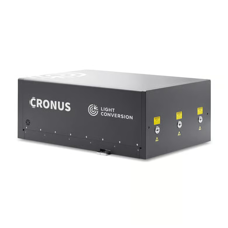
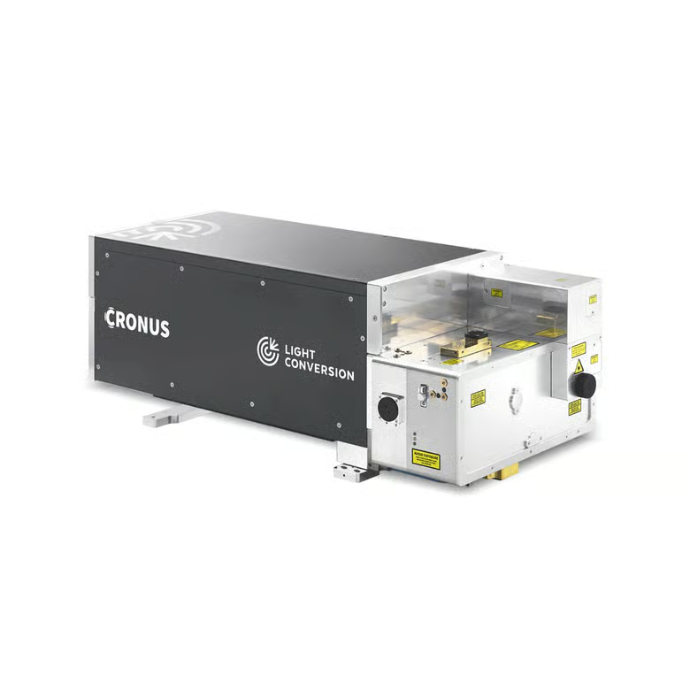
CRONUS-2P and CRONUS-3P are femtosecond sources built for nonlinear microscopy. They cover functional neuroimaging, optogenetics, and deep imaging using medium-repetition-rate three-photon excitation and fast high-repetition-rate two-photon imaging, as well as widefield and holographic excitation using high-power laser sources.
Zebrafish at 1300 nm
Multimodal 3D imaging of a live zebrafish at 1300 nm excitation using CRONUS-3P. 3PEF – green, trans SHG – yellow, epi THG – dark blue, trans THG – cyan. Courtesy of Luigi Bonacina group, University of Geneva.
Zebrafish at 1300 nm (2024)
Multimodal 3D imaging of a live zebrafish at 1300 nm excitation using CRONUS-3P. 3PEF – green, trans SHG – yellow, epi THG – dark blue, trans THG – cyan. Courtesy of Luigi Bonacina group, University of Geneva (2024).