Micro- and nanofabrication
Laser-tissue interaction
Femtosecond lasers offer exceptional precision and minimal thermal damage, making them suitable as treatment and surgical tools in medical disciplines such as ophthalmology.
Introduction
What laser–tissue interaction is
Laser–tissue interaction describes the processes that occur when laser radiation is absorbed by biological tissue. By selecting appropriate laser parameters and beam delivery conditions, laser irradiation can be tailored to achieve controlled tissue modification. Among the available laser technologies, femtosecond lasers are increasingly used for tissue processing because they enable highly localized energy deposition with limited thermal damage compared with longer-pulsed laser systems. These characteristics have enabled femtosecond lasers to be applied across a wide range of medical disciplines, including ophthalmology, dentistry, dermatology, and minimally invasive soft-tissue surgery.
Wavelength and tissue response
The interaction mechanism between biological tissue and laser irradiation depends on both the laser parameters – including wavelength, pulse duration, pulse energy, and repetition rate – and the properties of the irradiated tissue. These factors determine how laser energy is absorbed and distributed within the tissue, influencing the depth of the interaction and the resulting tissue response. UV radiation is strongly absorbed by biomolecules within superficial tissue layers and scattered by intracellular particles, resulting in shallow penetration depths that reach only tenths of micrometers. This makes UV lasers well suited for precise surface ablation but limits their use in procedures requiring access to deeper tissue targets. In contrast, near-IR femtosecond pulses can propagate through optically transparent tissues before being focused at a selected depth, enabling subsurface tissue modification without damaging the overlying layers.
How femtosecond pulses interact with tissue
Femtosecond laser ablation is based on depositing laser energy within the tissue on a timescale shorter than significant heat diffusion can occur. The high peak intensity of ultrashort pulses initiates nonlinear absorption and plasma formation within the irradiated volume. Once generated, the plasma absorbs the remaining laser energy and efficiently couples it to the tissue, producing a non-thermal ablation process that enables tissue resection with precise crater size, shape, and depth while minimizing thermal damage to the surrounding tissue. Besides laser fluence and repetition rate, scanning strategy plays an important role. Experimental studies have shown that appropriately designed scanning algorithms distribute laser energy while allowing efficient cooling between successive laser passes. By optimizing beam positioning and scan sequencing, local heat accumulation can be minimized even at high repetition rates.
Advantages of ultrashort pulses
The principal advantage of ultrashort pulses is the ability to remove tissue with high spatial precision while minimizing thermal damage to adjacent tissue. Compared with continuous-wave or longer-pulse lasers, ultrashort pulses provide precise tissue resection with minimal thermal damage and fine depth control, making them well suited for procedures where preservation of surrounding tissue is critical. Solid-state femtosecond lasers maintain good beam quality and high stability, while their ability to generate both IR and UV femtosecond pulses from a single laser platform enables multiple ophthalmic procedures to be performed using one laser source.
Ophthalmology
Ophthalmology represents one of the most established applications of laser–tissue interaction. Modern refractive surgery commonly combines two different laser sources: a near-IR laser for corneal flap creation and a UV laser for corneal stromal ablation. Near-IR pulses are focused within the transparent cornea, where localized photodisruption produces precisely positioned tissue separation without damaging the surrounding layers. UV radiation is strongly absorbed by corneal tissue and is therefore used for controlled surface ablation during photorefractive procedures. Recent studies have demonstrated that UV pulses generated by solid-state femtosecond lasers can achieve corneal stromal ablation comparable to, or in some cases superior to, conventional argon fluoride excimer lasers. High-speed transepithelial photorefractive keratectomy performed using 206 nm femtosecond pulses achieved ablation speeds of approximately 1.6 s per diopter while maintaining predictable and reproducible ablation together with healing responses comparable to excimer laser treatment. During the procedure, the increase in corneal surface temperature remained below 7 °C despite the high ablation speed. Solid-state femtosecond lasers generate UV radiation through harmonic generation, so both near-IR and UV pulses from the same laser source enable both intrastromal procedures and corneal surface ablation.
Dentistry
Dentistry is one of the emerging application areas for femtosecond laser tissue interaction, particularly in automated cavity preparation and restorative procedures. Robotic laser dentistry has been proposed as an alternative to conventional high-speed dental drills, enabling automated preparation of dental cavities while reducing the mechanical forces, vibration, and noise associated with rotary instruments. Studies investigating femtosecond laser ablation of dentin and enamel have demonstrated crack-free tissue processing without significant changes in chemical composition, tissue carbonization, melting, or deformation. Laser processing also avoids the formation of the dentin smear layer typically produced by high-speed mechanical grinding while preserving the morphology of the dentin structure and leaving dentinal tubules open. Compared with erbium and CO₂ dental lasers, which rely on water absorption and thermomechanical expulsion, femtosecond laser pulses remove tissue by ionizing the material directly into a plasma state. This process does not generate shock waves and produces superior surface quality; femtosecond lasers do not require water on the tissue surface and can process a wider range of restoration materials, including metals.
Dermatology
Dermatology represents another application area because many skin treatments require highly localized surface ablation. UV radiation is strongly absorbed within superficial tissue layers. Consequently, femtosecond UV lasers have been investigated as an alternative to nanosecond laser systems for dermatological procedures requiring controlled tissue removal with minimal collateral damage. Most biological effects of UV radiation below 320 nm are initiated when DNA absorbs light. Experimental studies comparing 206 nm and 257 nm femtosecond pulses have shown that the biological response depends strongly on wavelength, with 206 nm irradiation producing lower levels of DNA damage in skin cells than 257 nm irradiation under the investigated conditions.
Minimally invasive surgery
The high precision and limited thermal damage achieved with ultrashort laser pulses have also stimulated the development of minimally invasive surgical procedures. These characteristics are particularly important in gastrointestinal surgery, where colonic polypectomy and local excision require high precision together with minimal thermal damage because of the thin-walled structure of the bowel. Continuous-wave lasers, long-pulsed lasers, and electrocautery-based techniques are prone to significant thermal damage, with thermal damage to adjacent healthy tissue on the order of 300 µm in long-pulsed systems. Recent developments in hollow-core negative-curvature fibers have enabled high-energy ultrashort pulses to be delivered flexibly during endoscopic procedures. Preclinical studies using porcine colon tissue have demonstrated precise tissue resection using pulse energies of 46 and 33 μJ, with a thermally damaged region smaller than 85 μm together with fine control of ablation depth.
CARBIDE and PHAROS
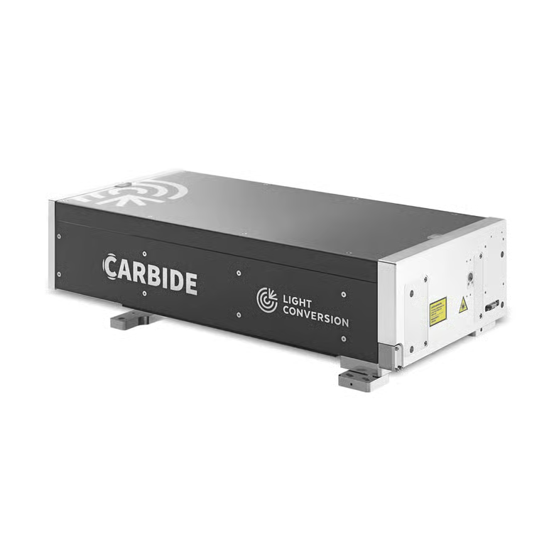
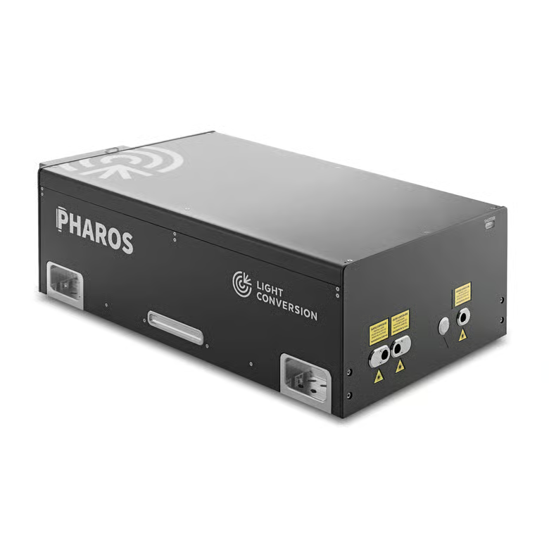
CARBIDE and PHAROS femtosecond lasers support a broad range of laser–tissue interaction applications by combining ultrashort pulse durations, high repetition rates, and integrated harmonic generation, providing output wavelengths from near-IR to UV. For ophthalmic applications, the fundamental near-IR femtosecond pulses can be used for intrastromal procedures, while UV pulses provided through harmonic generation can be applied for corneal surface ablation. The same wavelength flexibility supports dermatological procedures and other medical applications requiring precise tissue ablation.